Everything else

The rest of the job.

Conditions and feedback for patients and families, teaching and speaking, charity work, a few projects I'm proud of, media, and the trainee area.

Conditions

Developmental dysplasia of the hip: what parents ask me.

The questions I'm asked most often in the one-stop baby hip clinic we run at the Royal London every Wednesday afternoon, where clinical assessment, imaging and treatment all happen at the same time.

What is DDH?
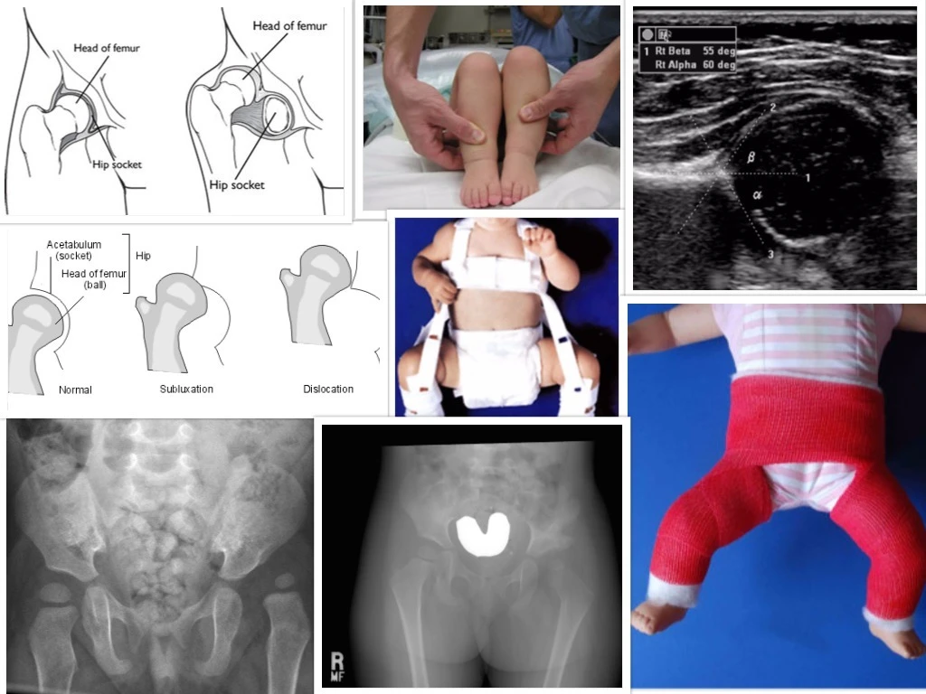
In DDH, the ball and socket of the hip joint do not fit snugly together, making the joint less stable, and the ball may slide in and out of the socket. This is called a subluxatable hip if the ball can pop back in. If the ball loses contact with the socket and stays outside the joint, it is called a dislocated hip.
How common is it?
DDH requiring treatment affects 1 to 3 babies per 1,000 births. That's up to a 1 in 300 chance of your child having this condition.
What are the risk factors?
The two most common are breech position (at any time during pregnancy or at birth) and a family history of DDH or hip problems in early life. Others include multiple pregnancy, oligohydramnios (low amniotic fluid), torticollis, and foot abnormalities such as positional talipes, metatarsus adductus and calcaneovalgus.
When should I suspect it?
When changing a nappy, one leg or thigh does not seem to open as fully as the other, or both seem restricted. Deep unequal creases in the buttocks or thighs. One leg appearing shorter than the other (bend both knees up and compare). A click or clunk when moving the hips. In older children, a limp, tiptoeing on the shorter leg, or a waddling gait if both hips are affected. Ortolani's and Barlow's tests are best performed by an experienced paediatric orthopaedic surgeon.
How is it diagnosed?
Often at the newborn and 6 to 8 week baby check. If not, see your GP, who will refer for an ultrasound scan (under 4 months of age) or an X-ray (over 4 months).
How is it treated?
Picked up under 4 months on ultrasound, treatment is a soft hip harness (Pavlik) holding the joint in position for 4 to 6 weeks, with a success rate above 90%. Later, or if the harness fails, the hip is gently relocated under anaesthetic (closed reduction) and held in a hip spica cast for 3 months. My preference is not to have a bar between the legs. In older children, further steps may be needed: open reduction, pelvic osteotomy (my preference is the Dega, which needs no pins or wires) and/or femoral osteotomy. I avoid leaving metalwork behind wherever possible, to prevent a second operation to remove it.
What complications should I be most concerned about?
Redislocation (most commonly within the first 10 days; treated by removing the cast and waiting until your child is older). Avascular necrosis, loss of blood supply to the hip, which is rare and often improves spontaneously if mild. The need for further surgery, either from loss of position or if the hip fails to develop fully as your child grows.
Conditions

Clubfoot: what parents ask me.

The questions I'm asked most often in the clubfoot clinic we run at the Royal London every Monday and Wednesday, where we provide comprehensive treatment for families of children born with clubfoot.

What is clubfoot?
Clubfoot, or talipes equinovarus, is a condition in which one or both feet are turned inwards and downwards at birth. Four elements are always present in some combination: cavus (high arch), adductus (forefoot turned in), varus (heel turned in) and equinus (foot pointing down). Some clubfeet are stiff and structural (idiopathic). Some are milder and correct with gentle stretching (positional). Both feet are affected in about half of babies with clubfoot.
How common is it?
Clubfoot affects roughly 1 in 1,000 babies born in the UK. That is about 700 babies each year in the country, and around 90 in London.
What are the risk factors?
Family history raises the risk. A couple with one affected child has a 3 to 5% chance of a subsequent child being affected. Male babies are affected roughly twice as often as female. Clubfoot can also appear as part of another condition (syndromic clubfoot), such as arthrogryposis or spina bifida, which is treated differently. The exact cause of most idiopathic clubfoot remains unknown, and nothing you did or did not do in pregnancy caused it.
How is it diagnosed?
Clubfoot is diagnosed at birth by the midwife or paediatrician on examination of the newborn. Once diagnosed, referral to a paediatric orthopaedic surgeon within the first few weeks of life is important, as treatment works best when started early.
How is it treated?
The gold standard is the Ponseti method, developed by Professor Ignacio Ponseti at the University of Iowa. Our Royal London team trained with his team in Iowa. Weekly gentle manipulation and casting corrects each of the four elements in sequence over 5 to 8 weeks. Around 90% of babies then need a small procedure called a percutaneous Achilles tenotomy to lengthen the tight heel cord, done under local anaesthetic. A final three-week cast is applied, and the baby then moves into the bracing phase. Extensive open surgery, once the norm, is rarely needed today, and I aim to avoid it wherever possible.
What is the boots and bar phase, and how long does it last?
The Foot Abduction Brace, known as "boots and bar", is two small boots joined by a bar that keeps the feet turned outwards to prevent relapse. It is worn 23 hours a day for the first 3 months, then only at night and during naps until around age 4. Brace compliance is the single strongest predictor of long-term success. Families who wear the brace as prescribed have a relapse rate below 10%. We spend time in clinic making sure every family is confident with the brace before leaving.
Will my child walk, run and play sport normally?
Yes, in the great majority. Children treated by the Ponseti method typically walk at the normal age, join in games with their peers at nursery, and play sport at school. Long-term studies of adults treated as babies show foot function, activity levels and quality of life close to those of the general population. The treated foot may be slightly smaller, and the calf slightly slimmer, on the affected side. Neither has functional consequences.
What complications should I be most concerned about?
Relapse is the most common issue, seen in 20 to 30% of children, most often between the ages of 1 and 5, and usually managed with a short course of re-casting. Anterior tibialis tendon transfer, a small operation in a preschool child, may be needed to hold the correction long-term. Skin problems under casts or the brace are rare with careful family training, which is a key part of our clinic. Long-term pain or stiffness in the corrected foot is uncommon.
Feedback

From patients and parents.

The feedback that counts more than anything else on this website.

"I truly believe that you could give somebody such awful news that they had two weeks to live and that person would leave your office removed of any fear or negativity, and this is really something that can't be studied in medical schools."

"My wife and I have both always agreed that as amazing and talented as every nurse, doctor, surgeon or consultant in the NHS is, they could learn so much more from a person like you."

Robert Walsh · father of Holly

"You'll be pleased to hear that Luke is walking completely unaided now. He is dancing, spinning around, bending his knees and trying to run, bless him. It's so amazing to see him wandering about on his own."

"His knee looks great, it's so good to finally see it bending in the right direction and the scar is so small. We are amazed and very thankful."

Colette and Torquil · parents of Luke

"My hip is perfect, I'm not having any issues with it at all. My rehab was good and I am hurdling without discomfort. You really have helped me with my dreams and for that I am truly grateful."

"I am currently ranked 4th in the country and not far from qualifying for the Olympic Games."

Richard Alleyne · athlete

"Jack is now three and a half. He truly is a remarkable little boy and nothing is holding him back, all thanks to the fantastic treatment he received from you in London."

"He loves being outside running, kicking the ball, playing on his bike, swimming. We will never forget everything you did for us, correcting Jack's feet so he can walk and run so well."

Linda Lawrance · mother of Jack, Australia

"Just a quick email to say that Sam is healing very well indeed after his recent op. We can't even find a scar."

Colin Blakemore · parent

"Thank you for your time and ongoing support with our son. We can only express how happy we are with the treatment you have provided to date."

Sean Drewery · father of Lewis
Teaching & speaking

Educational, keynote, judging and chairing gigs.
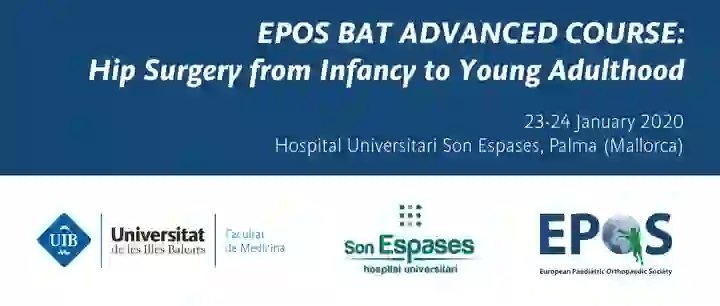
EPOS Paediatric Hip Course
Annual

EPOS Paediatric Hip Course

Palma, Mallorca & Geneva

Faculty on the European Paediatric Orthopaedic Society hip course.
Postgraduate Orthopaedic Courses, Singapore
Annual

Postgraduate Orthopaedic Courses

National University Hospital, Singapore

Faculty and organiser.

EPOS BAT Course
Annual

EPOS BAT Courses

Vienna, Austria

Basic and Advanced Traumatology instructional course trilogy.

Speaking topics

Charity work

Projects I've founded or been part of.
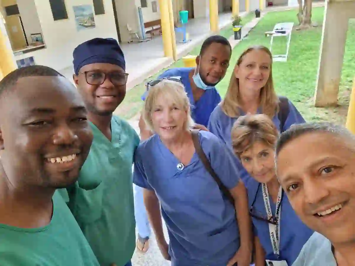
Mark Paterson Foundation, Ghana
2018–2025

Mark Paterson Foundation, Ghana

Co-founder of the Mark Paterson Trust, in association with MOTEC Life, in honour of my late colleague at the Royal London. Bi-annual trips of treatment and training to Ghana, where we see and treat complex children's bone and joint disorders in underserved populations.
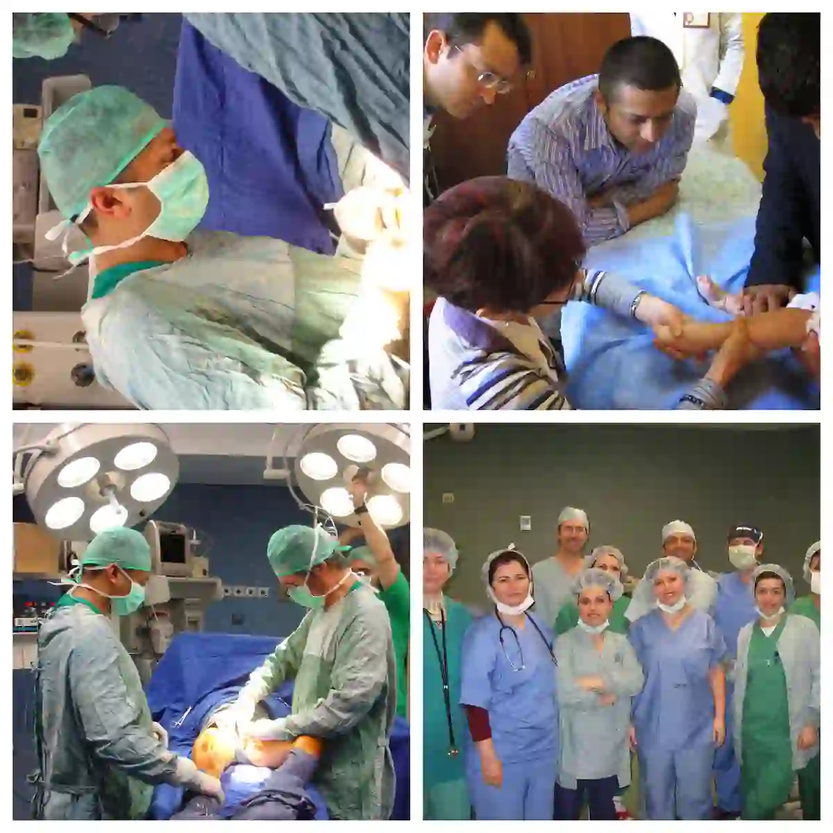
F.R.O.D.O., Albania
2009–2012

F.R.O.D.O., Albania

Foundation for Relief of Disabled Orphans. Assessing and treating children and young adults with bone and joint disorders alongside my colleague Mark Paterson. Five trips in total.

Some cool projects

A selection from the past.

PRINT study
Chief investigator · 2018–19

PRINT study

Innovate UK · £1.4m

The UK's first major study investigating 3D printed orthotics in children with cerebral palsy compared to standard orthotics.
Bespoke study
Chief investigator · 2015–17

Bespoke study

Urban cycling injuries · Barts Charity

A collaboration between the Orthopaedic and Emergency Departments of Barts Health to record the wheres, whys and hows of cycling-related injuries in London.
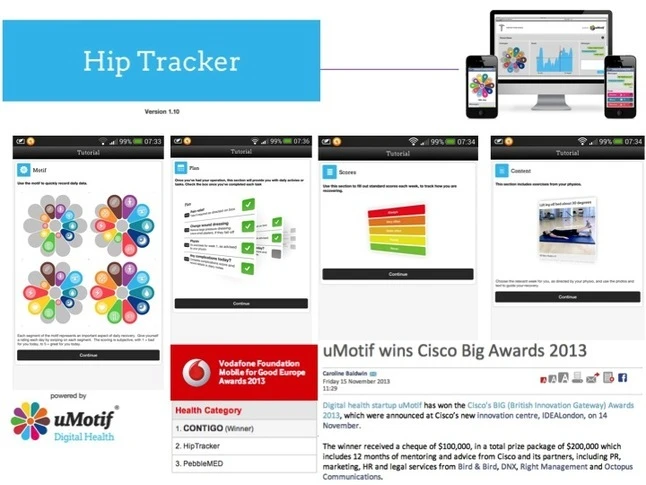
Hip Tracker
Chief investigator · 2013

Hip Tracker

with umotif

A smartphone app for patients after hip surgery to self-track symptoms and receive recovery content. Second place, Vodafone Foundation Mobile for Good Europe Awards 2013.
Walk This Way, Iowa
Organiser · 2009

Walk This Way

Barts & The London Charity

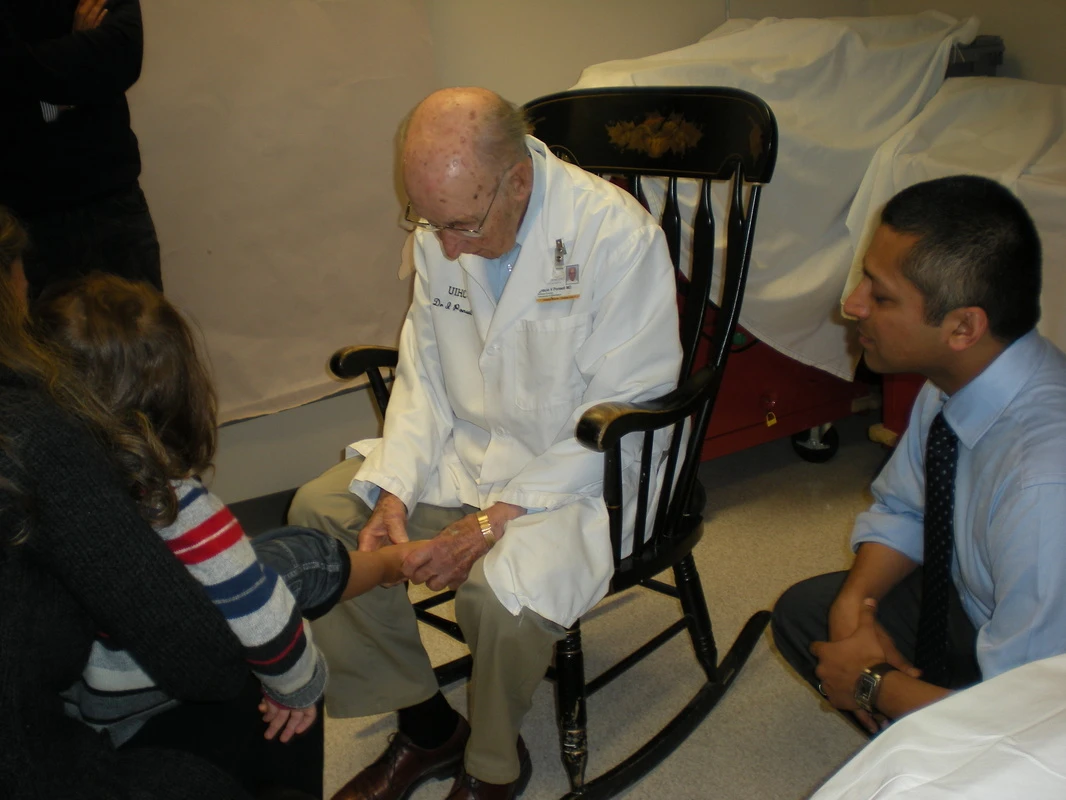
My team travelled to Iowa to learn the Ponseti method from Dr Ponseti himself. He died weeks after our visit. The Barts and The London children's foot clinic is now the UK's largest centre.
Children's Orthopaedic Outreach Programme
Chief investigator · 2015–17

Children's Orthopaedic Outreach Programme

Barts & The London Charity · £433k

A multidisciplinary programme bringing children's bone and joint care closer to home in North East London, with treatment initiated at Barts and virtual follow-up at home.

Media appearances

Some moments.

Save Our Squad, Disney+
Disney+ · 2022

Save Our Squad

Treating surgeon during David Beckham's Disney+ series, in which he mentored a London youth football team through their season.
BBC Inside Health, clubfoot
BBC Radio 4 · Feb 2016

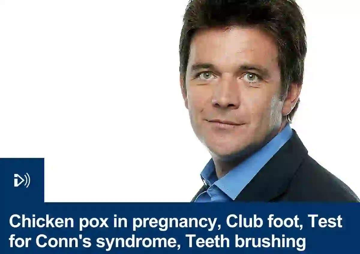
Inside Health: Clubfoot

Mark Porter visits the clubfoot clinic at the Royal London, which sent a team, led by me, to study with Dr Ponseti in Iowa.
BBC Inside Health, bandy legs and knock knees
BBC Radio 4 · Sep 2015

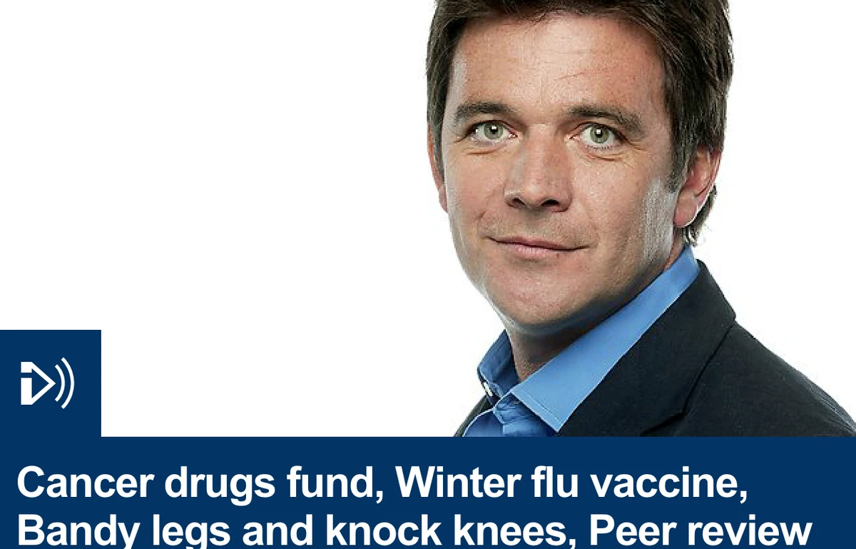
Inside Health: Bandy legs & knock knees

Up to a quarter of children referred to my clinics have normal developmental lower limb variants.
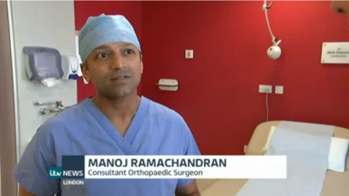
BMJ examination video
BMJ · Jul 2015

Normal lower limb variants in children

Clinical examination video recorded by the BMJ to accompany the review article.

Trainee area

Questions you need to know the answers to.

Viva questions drawn from Basic Orthopaedic Sciences, grouped by topic. If you can answer these, you are ready.

Statistics
Levels of evidence. Cohort versus case-control. Systematic review versus meta-analysis. Features of an RCT, bias, confounding, randomisation and blinding. Gaussian distribution, SD versus SEM, types of error, power analysis. Correlation and regression. Incidence versus prevalence. Screening programmes in orthopaedics and whether they meet the criteria. Contingency tables, sensitivity, specificity, PPV, NPV and accuracy. Reliability. Survival analysis. Funnel plots.
Genetics, embryology & pharmacology
Chromosome composition and DNA structure. Chromosomal abnormalities relevant to orthopaedics. Inheritance patterns and why they matter to surgeons. Osteogenesis imperfecta types and manifestations. When the limb bud first appears; secondary ossification centres; the layers of the physis and where fractures occur; rickets. Pain relief after arthroplasty; thromboprophylaxis for THA; bisphosphonates; drugs for osteoporosis; local anaesthetics and the haematoma block.
Inflammation, infection & imaging
Classifying bacteria; how penicillins act; resistance; pathogenesis of septic arthritis; MRSA; musculoskeletal TB; why prosthetic infections are hard to diagnose and treat. Plain radiography versus fluoroscopy; digital versus conventional; scatter and reducing exposure; reading a DEXA report; ultrasound and Doppler principles.
Tissue science: cartilage, bone, nerve, muscle, ligament, meniscus, disc
Composition and structure of articular cartilage and how structure relates to function; treatment options for cartilage defects. Structure of bone, its cells and matrix; calcium regulation; fracture healing and what affects it; bone grafts and allografts. Collagen structure; blood supply and healing of tendon and ligament; the stress–strain curve. Meniscal anatomy, vascular zones, function and repair. Nerve cross-section, classification of injury, axonotmesis, carpal tunnel, nerve conduction studies. Skeletal muscle structure, contraction, the neuromuscular junction, fibre types and healing. The intervertebral disc, annular tears and degeneration.
Biomechanics & biomaterials
Newton's laws in orthopaedics; force; moment; free-body diagrams and their assumptions. Stress–strain and Young's modulus; fatigue failure and S–n curves; plate thickness and rigidity; solid versus slotted nails; creep and uncemented stems. Bone cement; forging; femoral stem materials and coatings; highly cross-linked UHMWPE; the implant approval process. Biomechanics of the hip, knee, spine, shoulder, elbow, hand, wrist, foot and ankle.
Gait, prosthetics, orthotics & theatre
Prerequisites of normal gait; compensation for leg length discrepancy; eccentric and concentric gastrocnemius activity; the three rockers; causes of a positive Trendelenburg. What a prosthesis is, its indications, complications and what the surgeon must consider. Orthoses, how they work, materials, and the GRAFO. Theatre layout, sources of infection, ventilation systems, and moist bacterial strike-through.
Basic science of osteoarthritis
Defining OA and the fundamental principle of its pathogenesis. Biochemical changes in the extracellular matrix. Biomechanical changes in osteoarthritic cartilage. What an osteophyte is and how it forms. The biological mechanisms driving OA development.
Contact me

I would love to hear from you.

Clinical, teaching, speaking, research, media or venture enquiries.

Get in touch